Figure 1.
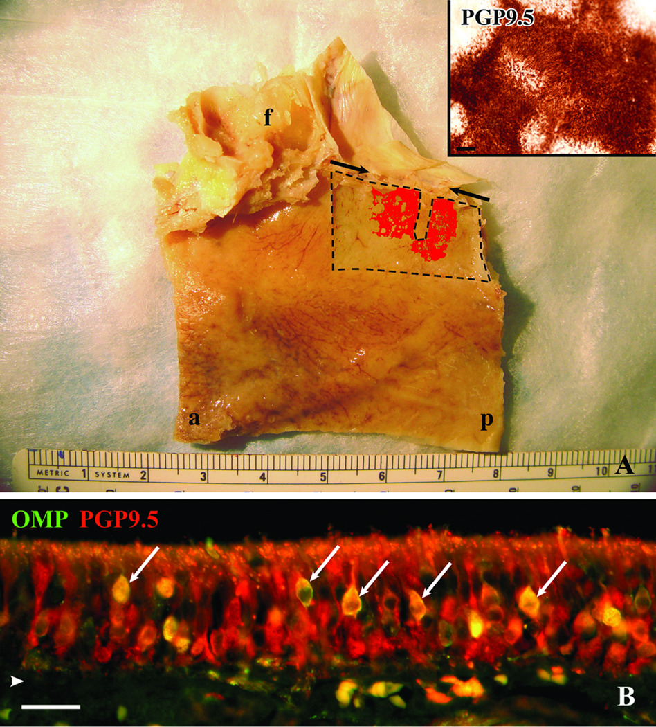
Autopsy specimens provide tissue for both a comprehensive perspective of the extent of neuronal staining and a detailed characterization of olfactory mucosa. A. Whole mount staining of the left septum taken from a 75 year-old male with Alzheimer’s. The edges of the whole mount stripped from the underlying bone are delineated by dashed lines. The thin rectangular defect extending from the superior boarder corresponds to the region removed from the whole mount for histology. The pattern of PGP9.5 staining of the mucosal whole mount is represented in red and is superimposed on a photograph taken of the septum after removal of the mucosa. The inset is a higher power magnification of the actual PGP9.5 staining at the surface of the whole mount. Notice the circular patches of non-neuronal epithelium at the edges of the olfactory boarder and within the olfactory region seen in the whole mount representation and inset. Arrows indicate anterior and posterior extent of the cribriform plate, f = frontal sinus, a = anterior, p = posterior, scale bar = 50µ. B. Section of epithelium from specimen shown in (A) fluorescently double labeled with OMP and PGP9.5 antibodies. PGP9.5 labels mature and immature olfactory neurons and OMP labels only the mature neurons. In this specimen the double labeled mature neurons (arrows) are relatively few compared to the abundant PGP9.5(+)/OMP(−) immature neurons. arrowhead = basement membrane, scale bar = 25µ.

