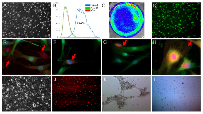
Figure 1. Characterization and differentiation of CPCs.
(A) The morphology of isolated CPCs growing on gelatin coated dish. (B) Flow cytometric analysis of purified Sca-1+ CPCs population. CPCs isolated from transgenic mice express robust (C) firefly luciferase and (D) GFP expression. After culturing in induction medium, differentiated CPCs stained positively with (E) connexin 43, (F) cardiac Troponin T, (G) myocyte-specific enhancer factor 2C, and (H) α-smooth muscle actin. (I) Cell morphology of CPC-derived endothelial cells. (J) These CPC-derived endothelial cells can uptake DiI-Ac-LDL. (K) These CPC-derived endothelial cells can form tube-like structure on Matrigel angiogenesis assay. (L) In contrast, the undifferentiated CPCs cannot form tube-like structure.

