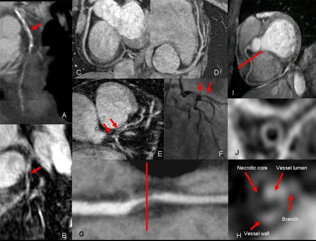
Figure 2.
a) Coronary CT of the left anterior descending coronary artery (LAD) showing severe calcification (red arrow) of the proximal and mid segment that obscure visualization of the lumen. However, corresponding b) coronary MR with contrast of the same LAD is not affected by calcium and reveals the area of critical narrowing (red arrow). High resolution coronary MR with 350 μm in plane resolution of the c) right coronary artery (RCA) and d) LAD. e) 350 μm coronary MR angiography showing severe narrowing of left main and LAD which is confirmed on f) conventional X-ray angiography. f) Multi-planer reformatted image of diseased RCA showing severe narrowing from non-calcified plaque, which demonstrates a g) necrotic core and enhancing wall on coronary NR with contrast. h) Coronary MR without contrast of normal RCA and i) corresponding black blood image showing the normal vessel wall of the mid segment.

