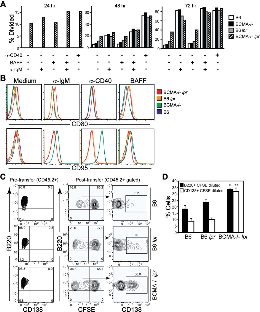
Figure 7. B cells from BCMA−/− lpr mice are hyperproliferative in vitro and in vivo.
(A) B cells (B220+ CD138−) isolated from spleens of 4-mo-old mice were labeled with CFSE and cultured 24–72 hrs in medium ± the indicated stimuli. Cells were recovered at each time point and the percent divided B cells were determined by flow cytometry. (B) Flow cytometric analyses of B cells analyzed in panel A after stimulation with the indicated stimuli for 72 hrs. Histograms of CD80 and CD95 expression levels are representative of 3–5 mice per genotype. (C) Donor B cells (B220+ CD138− CD3−) were purified from splenocytes of 2-mo-old B6, B6 lpr, and BCMA−/− lpr mice (all CD45.2+). Representative FACS plots demonstrating that the purity of donor B cells before transfer was >95% are shown. Cells were CFSE-labeled and 15 × 106 were injected i.v. into CD45.1+ wild-type recipients. 3 d after transfer, the percentage of dividing CD45.2+ B220+ B cells and differentiation into CD138+ PCs in the spleens of recipient mice were measured by flow cytometry. (D) Data are expressed as means ± SEM (n = 6 recipients per donor group). *, p < 0.05; **, p < 0.01 compared to B6 and B6 lpr donor groups.

