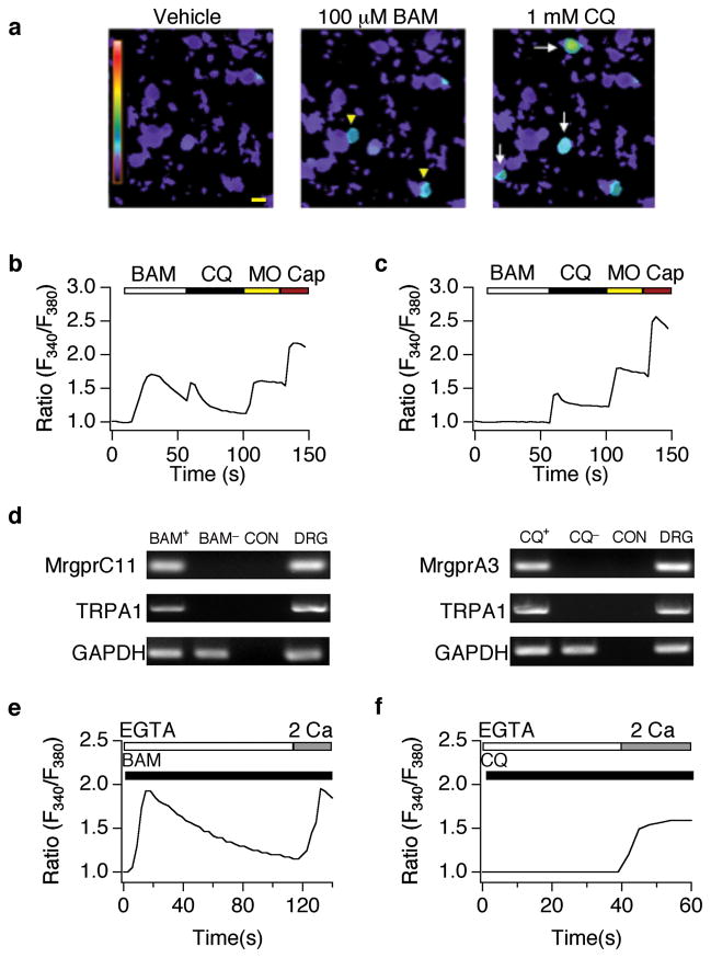
Figure 1. Chloroquine and BAM8–22 activate a subset of TRPA1-positive sensory neurons.
(a) BAM8–22- (BAM; 100 μM; yellow arrowheads) and chloroquine (CQ; 1 mM; white arrows)-evoked responses in cultured dorsal root ganglia neurons (representative Fura-2 ratiometric images). Scale bar=10 μm. (b) Representative BAM- and CQ-responsive cell. Fura-2 ratio in response to BAM (100 μM), CQ (1 mM), allyl isothiocyanate (mustard oil: MO; 200 μM), and capsaicin (Cap; 1 μM). (c) Representative CQ-sensitive, BAM-insensitive cell. Fura-2 ratio in response to BAM (100 μM), CQ (1 mM), MO (200 μM), and Cap (1 μM). (d) PCR analysis of MrgprA3, MrgprC11, and TRPA1 expression in CQ-positive, BAM-positive and CQ/BAM/MO-negative large diameter sensory neurons. MrgprC11 and TRPA1 were amplified in BAM-sensitive (BAM+), but not BAM-negative (BAM−) or no-RT control (CON) cells (right). MrgprA3 and TRPA1 were amplified in chloroquine-positive cells (CQ+), but not chloroquine-negative (CQ−), or no-RT control (CON) cells (left). MrgprA3, MrgprC11 and TRPA1 were all amplified from DRG cDNA (DRG). Note the presence of control GAPDH product in all samples. (e) Representative trace showing Ca2+ response to BAM (100 μM) in the absence (1mM EGTA), and presence (2 mM Ca2+) of extracellular calcium. (f) Representative response to CQ (1 mM) in the absence (1 mM EGTA), and presence (2 mM) of extracellular calcium.

