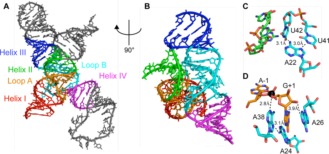
Figure 8.
(A) Crystal structure of a 4WJ construct with a vanadate transition state mimic(9), color coded by helices I-IV and loop A and B. (B) Rotated view emphasizing docking interface between loop B and helix II/loop A. (C) Involvement of A22 in orienting U42 at the loop B/helix II interface. (D) Adenosine residues in vicinity of G(+1) and the vanadate transition state mimic (black ball).

