
Fig 6. Immunoreactivity of HDACs in primary lymphoma tissue sections. A) diffuse large cell lymphoma.
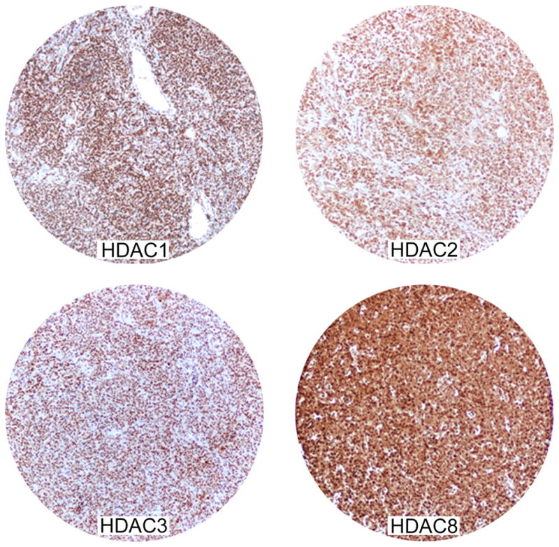

Most tumour cells stained positive for HDAC1,2,3 and 8 in all cases. The staining for HDAC1, 2, and 3 is nuclear with a strong intensity; the staining for HDAC8 was either nuclear or cytoplasmic with strong intensity. B) classical Hodgkin lymphoma. HRS cells (arrow) and the surrounding reactive cells expressed HDAC1, 2, 3, 8 and 10, but not HDAC6.