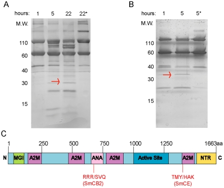
Figure 3. Human complement C3 is cleaved by both SmCE and SmCB2.
In vitro cleavage of human complement C3 protein confirms ex vivo analysis, and reveals differential cleavage by CE and CB2. (A) SmCB2 digestion of complement C3. (B) SmCE digestion of complement C3. Digestion reactions were performed for 1–22 hours (SmCB2) or 1–5 hours (SmCE) at 37°C. * indicates pre-incubation with (A) 1 mM CAO74 or (B) 1 mM AAPF-CMK. ** indicates no peptidase control. Arrows indicate bands submitted for Edman degradation. (C) Schematic of human complement C3, indicating cleavage sites as determined by Edman degradation (MGI-macroglobulin-1; A2M-alpha-2-macroglobulin; ANA-.Anaphylatoxin homologous domain; NTR-netrin-like domain).

