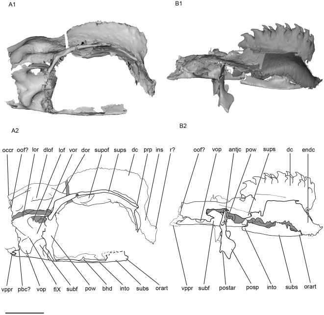
Figure 3. Lateral view of the skull of Kawichthys.
Right side. A1, surface rendering generated from Synchrotron Radiation X-ray microtomographic slices of KUVP 56340. Part of the dorsal surface of the interorbital plate removed (dotted line). A2, corresponding drawing. B1, surface rendering generated from Synchrotron slices KUVP 152144. Dark grey, internal surface of the endocranial cavity and dermal median dorsal crest; light grey, external surface of the braincase. B2, corresponding drawing. Scale bar = 0.5 cm. Abbreviations: antjc, anterior opening of the jugular canal; bhd, bucco-hypophyseal duct; dc, dorsal crest; dlof, dorsolateral otic fossa; dor, dorsal otic ridge; endc, endocranial cavity; fIX, foramen for the glossopharygeus nerve; ins, internasal septum; into; interorbital plate; lof, lateral otic fossa; lor, lateral otic ridge; orart, orbital articulation for the palatoquadrate; occr, occipital crest; oof?, otico-occipital fissure; pbc?, posterior basicapsular commissure; postar, postorbital articulation for palatoquadrate; postp, postorbital process; pow, postorbital wall; prp, preorbital process; r?, rostrum; subf, subotic fossa; subs, suborbital shelf; supof, suborbital fenestra; sups, supraorbital shelf; vop, ventral otic process; vor, ventrolateral otic ridge; vppr, ventral paroccipital process.

