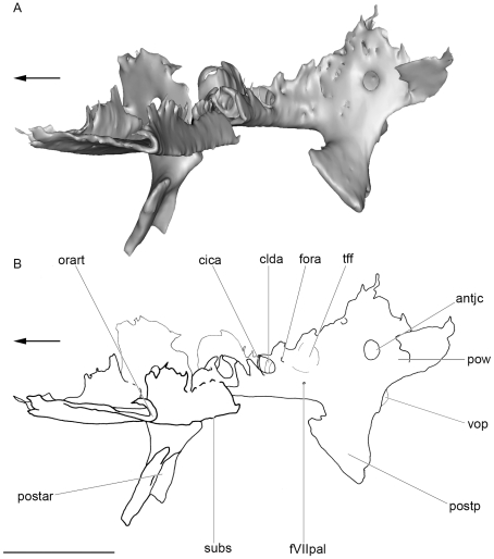
Figure 10. Oblique medial view of the basicranium of KUVP 152144.
Part of the dorsal surface of the interorbital plate removed (dotted line). Left side. A, surface rendering generated from Synchrotron Radiation X-ray microtomographic slices. B, corresponding drawing. Arrows points forward. Scale bar = 0.5 cm. Abbreviations: tff, trigemino-facial fossa. See Figs. 3, 4, 6 and 7 for other abbreviation.

