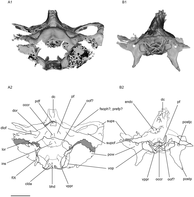
Figure 11. Posterior view of the skull of Kawichthys.
A1, surface rendering generated from Synchrotron Radiation X-ray microtomographic slices of KUVP 56340 (A). A2, corresponding drawing. B1, surface rendering generated from Synchrotron Radiation X-ray microtomographic slices of KUVP 152144. Dark grey, internal surface of the endocranial cavity and dermal median dorsal crest; light grey, external surface of the braincase. B2, corresponding drawing. Scale bar = 0.5 cm. See Figs. 3, 4, 5 and 6 for the abbreviations.

