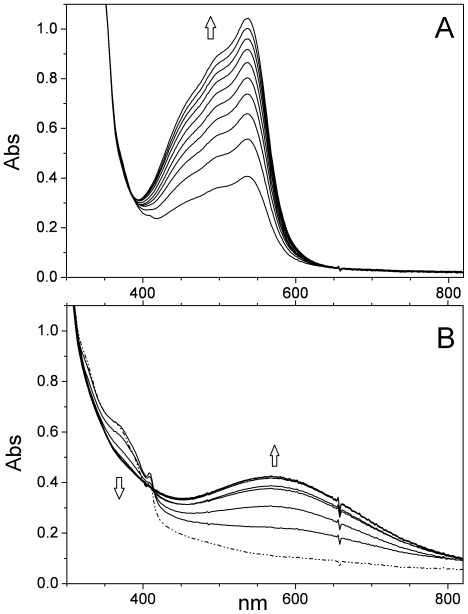
Figure 8. Reaction of reconstituted EBV R2 with scavengers.
(A) Representative light absorption spectra recorded at T = 288 K of reconstituted EBV R2 (∼130 µM, pH 7.5, 50 mM HEPES buffer) after addition of 1 mM bathophenanthrolinesulphonate and 4 mM HU, recorded in 10 min intervals under anaerobic conditions. (B) Reconstituted EBV R2 (∼130 µM) incubated with 1 mM catechol at pH 7.5 under anaerobic conditions at T = 293 K. The light absorption spectra were recorded at 30 s intervals. The dashed-dotted line represents the spectrum of the reconstituted diferric protein without any metal-ion chelator.

