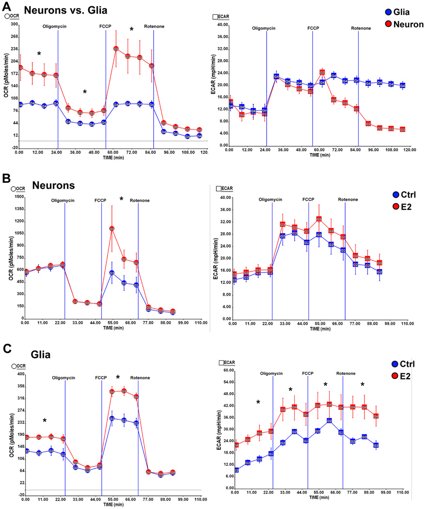
Figure 6. Estrogen differentially regulates mitochondrial bioenergetics in neurons and glia.
Primary hippocampal neurons from day 18 (E18) embryos of female Sprague-Dawley rats were cultured in Neurobasal medium + B27 supplement for 10 days prior to experiment. Mixed glia from day 18 (E18) embryos of female Sprague-Dawley rats were cultured in growth media (DMEM:F12 (1:1)+10% FBS). Primary Oxygen consumption rate (OCR) and extracellular acidification rate (ECAR) were determined using the Seahorse XF-24 Metabolic Flux analyzer. Vertical lines indicate time of addition of mitochondrial inhibitors A: oligomycin (1 µM), B: FCCP (1 µM) or C: rotenone (1 µM). A, left panel: higher basal OCR rates and larger maximal mitochondrial respiratory capacity in primary neurons (red) than mixed glia (blue); right panel: comparable glycolysis rate (indicated by ECAR) between neurons (red) and mixed glia (Blue) (*, P<0.05 compared to mixed glia, n=5 wells per group); B, left panel: E2 treatment (red) increased the maximal mitochondrial respiratory capacity but not the basal respiration in neurons; right panel: E2 treatment did not significantly increase glycolysis (indicated by ECAR) in neurons (*, P<0.05 compared to control (blue), n=5 wells per group); C, left panel: E2 treatment (red) significantly increased both the basal respiration and the maximal mitochondrial respiratory capacity in mixed glia; right panel: E2 treatment (red) significantly increased glycolysis (indicated by ECAR) in mixed glia (*, P<0.05 compared to control (blue), n=5 wells per group).

