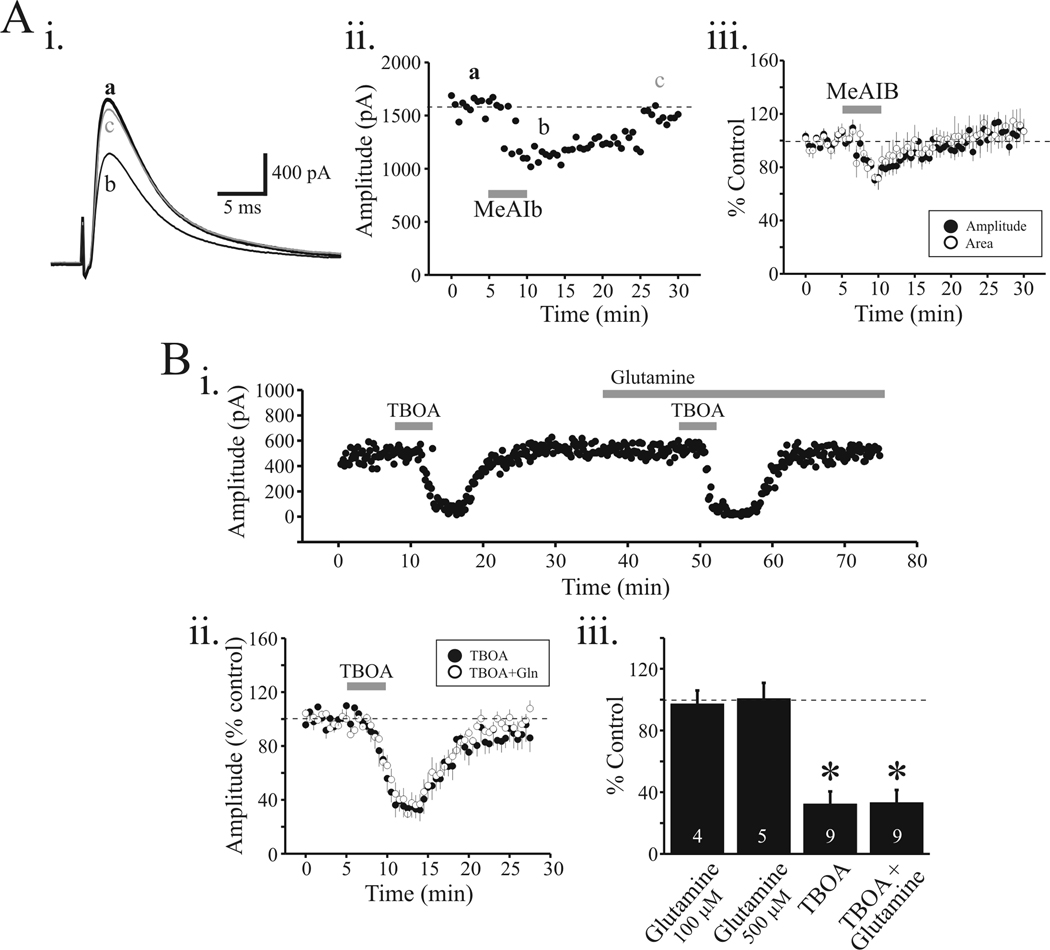
Figure 2.
MeAIB and TBOA reduced eIPSC in isolated VB slice. Ai. In this preparation, the TRN was dissected from the slice. Representative recording from VB neuron and the eIPSC evoked by local electrical stimulation in control (a), following MeAIB application (10 mM) (b) and 15 min wash (c). Note the decrease in amplitude of eIPSC following MeAIB application. Aii. Time course of MeAIB-mediated action on eIPSC. Aiii. Population data indicating the reduction of eIPSC amplitude (closed circles) and eIPSC area (open circles) by MeAIB. Bi. In a different relay neuron, TBOA strongly attenuates the eIPSC that recovers to baseline levels within 10 minutes. In the presence of glutamine (100 µM, 500 µM), the reduction in eIPSC by TBOA is unaltered. Bii. Population data indicate similar decreases in eIPSC amplitude by TBOA before (filled circles) and in the presence of glutamine (open circles). Biii. The histogram plot summarized the average change in eIPSC amplitude by glutamine alone (100 µM, 500 µM), TBOA alone, and TBOA + glutamine. Data are presented as percent of control levels. * p<0.01.

