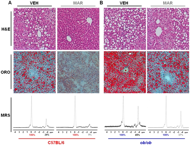
Figure 3. Marimastat reversed hepatic steatosis in diet-induced obese mice as well as in ob/ob animals, as demonstrated by histology and magnetic resonance spectroscopy.
Representative liver sections stained with hematoxylin and eosin (H&E; top panels; original magnification 200×), and Oil Red O (ORO; middle panels; original magnification 200×). Hepatic fat fraction was measured by magnetic resonance spectroscopy (MRS; bottom panels, representative animals). Percent fat content was determined relative to water (100%) by numerical integration of the areas under the lipid and water peaks. Livers from MAR treated diet-induced obese animals exhibited almost normal hepatic architecture, whereas VEH livers revealed micro- and macrovesicular steatosis (A). Livers from VEH treated ob/ob mice revealed extensive, predominantly macrovesicular steatosis (B). MAR treated livers from ob/ob mice, in contrast, showed a decrease in macrovesicular lipid droplets mainly around the portal tract (B). Histological findings were confirmed with MRS, demonstrating a decrease in fat content in Marimastat-treated animals (A,B bottom panels).

