
Figure 5.
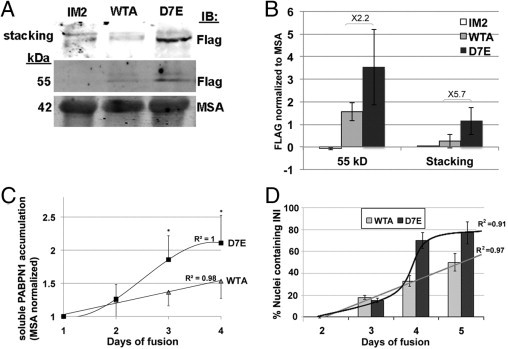
expPABPN1 aggregation potency is higher than WT-PABPN1 in myotubes. A: Western blot analysis of total protein extracts isolated from IM2, WTA, and D7E myotubes. PABPN1-FLAG is detected with an anti-FLAG antibody. Aggregated PABPN1-FLAG is retained in the stacking gel, and the soluble protein migrates at 53 kDa. Muscle skeletal actin (MSA) is used as a loading control. B: Quantification of PABPN1-FLAG accumulation in IM2, WTA, and D7E myotubes at 4 days after fusion. The histogram shows the FLAG signal after normalizing to actin. Averages are calculated from six independent experiments. C: Quantification of soluble PABPN1-FLAG protein accumulation in WTA and D7E during cell fusion. Values show the FLAG signal normalized to MSA. Averages are calculated from six replicates. *P < 0.05. Linear or sigmoid curve fits of PABPN1-FLAG accumulation in WTA or D7E, respectively (R2 values are indicated). D: INI formation in WTA and D7E cell lines during myoblast cell fusion. INIs were visualized with an anti-FLAG antibody. Histograms show the percentage of nuclei containing PABPN1 INI from 80 to 100, counted in 20 myotubes at 2, 3, 4, and 5 days after fusion. A linear or sigmoid curve fits INI formation in WTA or D7E, respectively (R2 values are indicated).