
Figure 2.
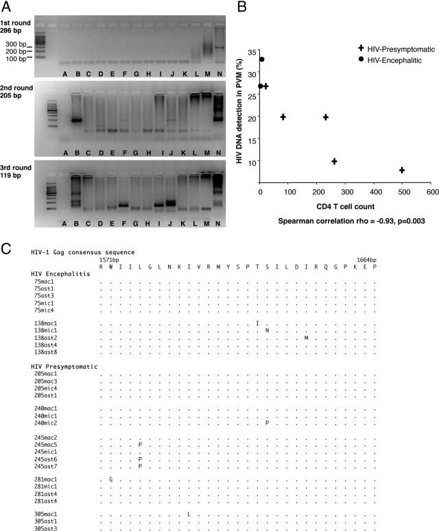
Analysis of HIV-1 gag DNA isolated from laser microdissected brain cell populations; astrocytes, perivascular macrophages (PVM), and parenchymal microglia (PM) from HIV-presymptomatic (PS) and HIV-encephalitic (HIVE) cases. A: Representative images of HIV-1 gag DNA PCR resulting in three rounds of PCR products (286, 205, and 119 bp). HIV-1 gag DNA was not consistently detected in PCR amplification from samples in which there were low levels of virus. Cell populations were therefore collected in duplicate (two collections of astrocytes per case), and the triple-nested PCR was performed on each cell population at least 10 times, to increase the frequency of detection. Not every PCR amplification from DNA of cell populations is illustrated (results of PCR amplifications of HIV-1 gag DNA from all cell populations are given in Table 1 and sequencing results are shown here in panel B). Lane A, negative PCR control (water as DNA template) carried through three rounds of PCR. Lane B, code no. 281 (PS), PVM. Lane C, code no. 305 (PS), PVM. Lane D, code no. 281, astrocytes. Lane E, code no. 305, astrocytes. Lane F, code no. 75 (HIVE), astrocytes. Lane G, code no. 75, PVM. Lane H, code no. 138 (HIVE), astrocytes. Lane I, code no. 138, PVM. Lane J, code no. 75, PM. Lane K, code no. 138, PM. Lane L, code no. 281, PM. Lane M, code no. 305, PM. Lane N, Lymph node of HIV-positive patient, positive PCR control (overamplification results in PCR product not running through the gel). B: Association between CD4 T-cell count and the frequency with which HIV-1 DNA was detected by triple-nested PCR in perivascular macrophages (PVM, expressed as a percentage) in the occipital cortex for each of the seven HIV-positive individuals. C: Amino acid sequence alignment of HIV-1 gag DNA from PCR products of laser microdissected brain cell populations. Sequences are aligned and numbered according to the HIV-1 gag consensus sequence. ast, astrocyte; mac, perivascular macrophages; mic, parenchymal microglia.