
Figure 3.
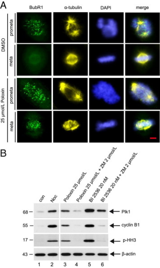
Poloxin activates the mitotic checkpoint. A: HeLa cells were synchronized by thymidine treatment and released into medium containing DMSO or 25 μmol/L Poloxin for 10 hours. Cells were stained for BubR1, α-tubulin, and DNA. Scale bar = 5 μm. B: HeLa cells were synchronized by thymidine treatment and released into medium containing 25 μmol/L Poloxin or 20 nmol/L BI 2536. Twelve hours later, the shake-off cells were further incubated with Poloxin alone, Poloxin plus 2 μmol/L ZM 447439, BI 2536 alone, or BI 2536 plus ZM 447439 for a further 2 hours, as indicated. Cellular extracts were then prepared for Western blot analyses with antibodies against Plk1, cyclin B1, and p-HH3. Nontreated (con) or nocodazole-treated (Noc) cells were taken as mitotic-negative and mitotic-positive controls, respectively. β-Actin served as the loading control.