
Figure 2.
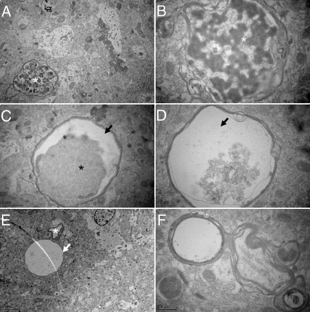
Axonal disease. Ultrastructural aspects of axonal degeneration representing various stages. A: Accumulation of autophagic vacuoles (asterisk). B: Example of digested axoplasm (asterisk). C and D: Accumulation and detachment (arrows) of dark axoplasm (asterisk). E: Extremely swollen degenerated axon is almost completely electron-lucent (arrow) next to a capillary (asterisk). F: Axon with split myelin sheath.