
Figure 5.
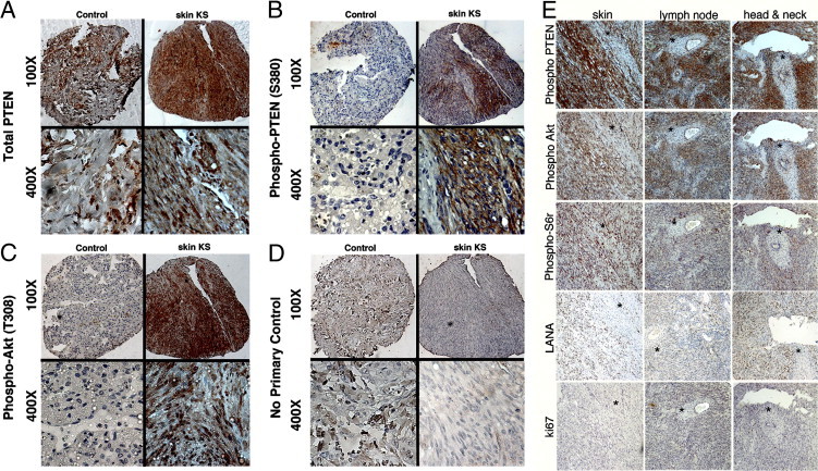
Expression of PI3K activation markers and phospho-PTEN (S380) in KS primary biopsies. Immunohistochemistry showed the presence of total PTEN (A), phospho-PTEN (S380) (B), phospho-Akt (T308), a PI3K-dependent site (C), and no-primary-antibody negative control (D). Original magnification: ×100 (upper row); ×400 (lower row). In each panel (A–D), images at the left are of control lung tissue, which stains positive only for PTEN but not for phospho-PTEN (S380) or phospho-Akt (T308); images at the right are of a representative skin biopsy, in which the same tissue section is positive for total PTEN, phospho-PTEN (S380), and phospho-Akt (T308). E: Staining of a second TMA incorporating AIDS-KS from tissues of three different origins for phospho-PTEN (S380), PI3K activation markers phospho-Akt (T308) and phospho-S6r (S235/236), KSHV marker LANA, and proliferation marker Ki67. Sections were selected such that a region of the biopsy was negative (asterisks), in an otherwise positive section, serving as an internal control. Original magnification, ×100.