
Figure 3.
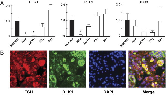
Paternally expressed genes. A: Expression of DLK1, RTL1, and DIO3 in clinically NFA, ACTH-secreting, PRL-secreting, and GH-secreting pituitary adenomas was detected by quantitative real-time PCR as described under Materials and Methods. Values from normal pituitaries were designated as 1, against which values from pituitary tumors were normalized. Data are reported as means ± SEM. Student's t-test was used to compare values between tumors and the normal pituitaries. *P < 0.05. B: Coexpression of DLK1 and FSHβ in normal human anterior pituitary. Sections of normal pituitaries were immunostained with antibodies against DLK1 and FSHβ, and nuclei were stained by DAPI. The merged image demonstrates the co-localization (yellow) of DLK1 and FSHβ in normal pituitary cells. Original magnification, 400×.