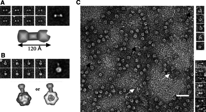
Fig. 3.
Visualisation of Ad L4-100 K protein alone and in the complex with hexon. a Gallery of eight EM images of the recombinant Ad2 L4-100 K protein, the averaged image, and the structural model of the L4-100 K protein (from left to right, upper panel). The L4-100 K protein is a dumbbell-shaped molecule, consisting of two globular domains linked by a rod-like structure. The total length of the molecule is about 120 Å. b Gallery, the averaged image and the model of the recombinant Ad3 hexon-Ad2 L4-100 K protein complex, with the hexon in end-on view (from left to right, lower panel). One of the globular domains of the L4-100 K protein is attached either to the distal domain or to the apical domain of the hexon trimer. c EM analysis of native proteins purified from Ad5-infected 293 cells. Scale bar 30 nm. Most of the visible structures are hexons. Some L4-100 K protein molecules and hexon–100 K protein complexes are marked with black and white arrows, respectively. The column on the right represents five images of hexon–100 K protein complexes and five images of 100 K protein molecules (upper and lower columns, respectively) (from [94])

