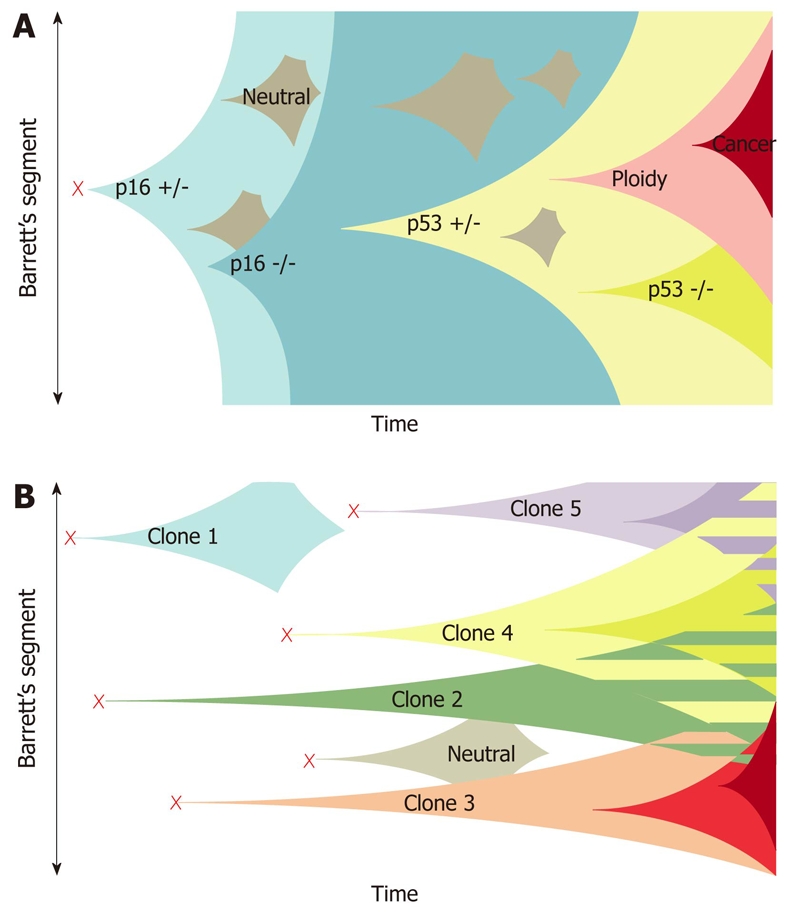
Figure 1.

Clonal evolution models in Barrett’s esophagus. A: The current model of clonal evolution adapted from Maley et al[66]. Founder mutation (red cross) occurs in a single progenitor and provides a growth advantage that predisposes to a selective sweep. Successive selective sweeps result in progression along the metaplasia dysplasia pathway. Clone bifurcation is responsible for the genetic heterogeneity in this model; B: The newly proposed model of evolution based on the mutation of multiple progenitor cells situated in esophageal gland squamous ducts located throughout the length of the esophagus (red crosses). Multiple independent clones then arise and evolve separately. The presence of multiple different clones gives rise to a mosaic interdigitating clonal pattern of the Barrett’s segment represented as the striped areas[65].
