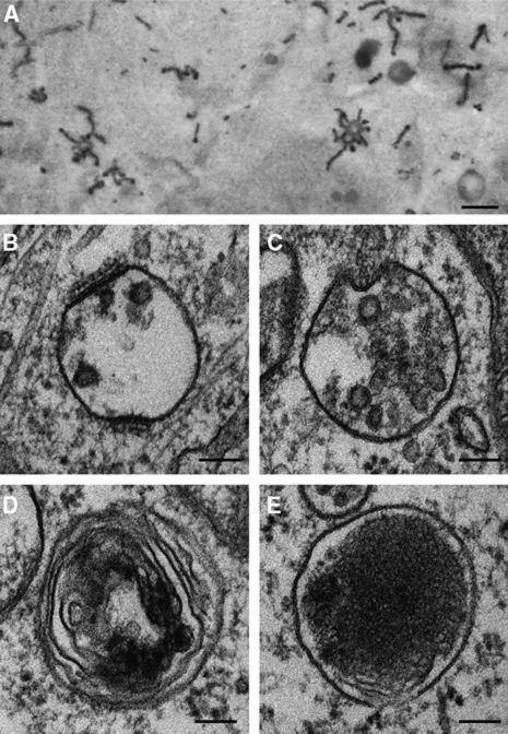
Figure 3.
Morphologies of endosomes and lysosomes at the ultrastructural level. (A) Electron micrographs of peripherally located EEs containing HRP-conjugated Tf. They contain vacuolar and tubular domains. Courtesy of Tooze and Hollinshead (1992). Electron micrographs of (B) EE with clathrin lattices and a few ILVs; (C) LE, containing numerous ILVs; (D) endolysosome, with partial electron dense areas; and (E) lysosomes, with electron dense lumen. Images are all from HeLa cells that had been processed for thin section EM. Scale bars in (A): 500 nm and (B–E): 100 nm. Figure 3A is reproduced with kind permission from Rockefeller University Press;©2009 Rockefeller University Press. Originally published in J Cell Biol 118: 813–830. doi: 10.1083/jcb.118.4.813.

