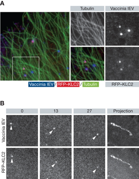
Figure 3.
Vaccinia IEV recruit kinesin-1 and move on microtubules. (A) Immunofluorescence images showing recruitment of kinesin-1 (RFP–KLC, red) to vaccinia IEV (blue) associated with microtubules (green). (B) Stills taken from live cell imaging of the movement of YFP-tagged vaccinia IEV in cells expressing RFP-tagged kinesin light chain 2. The time in seconds is indicated and the right panel shows a maximum intensity projection to highlight the path taken by the virus.

