
Figure 2.


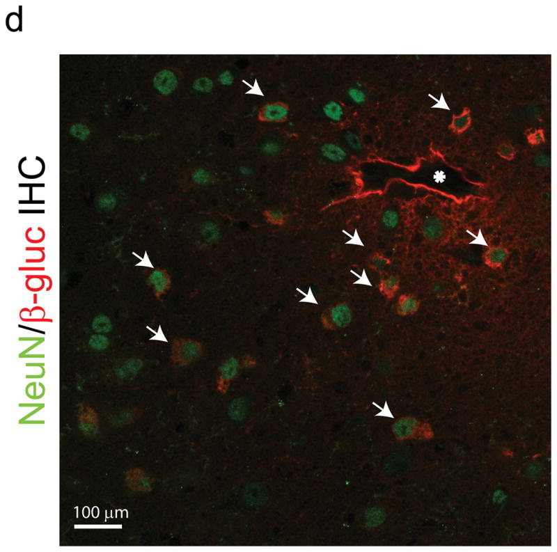
Peptide epitopes expand the tropism of AAV2. (a,b) Viral genomes in brain and liver of wildtype (a) and MPS VII (b) mice as quantified by Q-PCR after peripheral injection. Data are mean ± SEM. (a) AAV-PPS and AAV-TLH are significantly enriched in brain (p<0.001, students t-test) vs. AAV-WT. (b) AAV-PFG and AAV-LSS are significantly enriched in brain over AAV-WT (p<0.001, student’s t-test). (c) AAV-PFG.βgluc transduces the cerebral vasculature of MPS VII mice after tail vein injection. Representative sections were stained for β-glucuronidase activity in situ, which leaves a red precipitate. Representative photomicrographs from sections show that vessels throughout the brain display activity when MPS VII mice (left and middle panels), but not wildtype mice (upper right), were injected. (d) Confocal microscopy of sections stained with anti-β-glucuronidase (red) and anti-NeuN (green) reveals vascular and neuronal β-glucuronidase staining, respectively (denoted by arrows).