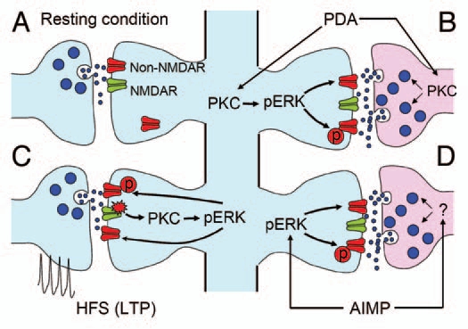
Figure 1.

Schematic diagram summarizing PBA-CeAC synaptic transmission in normal and AIMP mice. (A) Synaptic transmission in a resting condition at PBA-CeAC synapses is mediated by non-NMDARs. (B) Application of PDA activates PKC in presynaptic terminals and postsynaptic dendrites. Activated PKC in presynaptic terminals enhances glutamate release, while that activated in postsynaptic spines activates ERK to upregulate non-NMDAR functions or increase their numbers at synaptic sites. (C) High-frequency stimulation activates NMDARs, which in turn activate the PKC-ERK signal pathway to upregulate non-NMDAR functions or increase their numbers at synaptic sites, thereby resulting in LTP. (D) In mice with AIMP, ERK is activated in the CeAC by an excessive nociceptive signal, which in turn upregulates non-NMDAR functions or increases their numbers at synaptic sites. In addition, enhanced glutamate release from presynaptic terminals was also observed in AIMP mice. Together, these pre- and postsynaptic enhancements at PBA-CeAC synapses might partially account for the central sensitization in AIMP.
