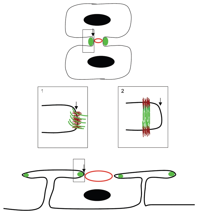
Figure 1.

Hemicentins assemble in the cleavage furrow. Schematic diagram of hemicentin (green) in the incomplete cleavage furrow of a conventional cell (e.g., mouse blastomere, top) and a syncytial germ cell of the C. elegans gonad (bottom). An expanded view (box 1) shows a simple model in which hemicentin (green) assembles as an elastic ring around the periphery of the cleavage furrow leading edge. An unconventional model (box 2) shows hemicentin in a distal position from the leading edge of the cleavage furrow. Hemicentin polymers may be assembled perpendicular to the membrane, linking the two cellular products of cytokinesis, a distribution that is more consistent with the role of hemicentins as elastic connectors between somatic cells in C. elegans. Also shown are nuclei (black circles) contractile rings (red circles) and putative transmembrane receptors (brown lines).
