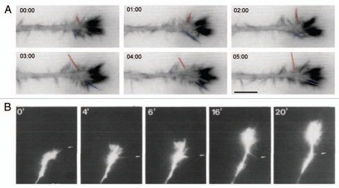
Figure 2.
Comparison of growth cone dynamics on the line substrate and in a developing retina. (A) Growth cone dynamics of N1E-115 neuron-like cells on the line substrate. N1E-115 were transfected with GFP-lifeact, an F-actin marker. Red and blue lines indicates the lateral movement of non-aligned filopodia. Note the high F-actin content in aligned filopodia. Timescale is in minutes:seconds. Part reproduced with permission from Jang et al.5 (B) Growth cone dynamics of Xenopus retinal axons imaged live in the developping retina. Note streamlined appearance of growth cone and left-right lateral movement of lateral filopodia (indicated by arrows). Part reproduced with permission from.12 Scale bars = 10 mm.

