. 2001 Dec;3(4):265–279. doi: 10.31887/DCNS.2001.3.4/fxvollenweider

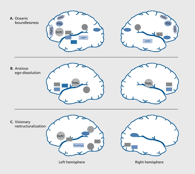
Figure 4. Correlations between the three dimensions of the APZ questionnaire for altered states of consciousness (oceanic boundlessness [OB], anxious ego-dissolution [AED], and visionary restructuralization [VR]) and regional brain activity (cerebral metabolic rate of glucose [CMRglu]) in healthy volunteers under psilocybin (0.26 mg/kg PO)orS-ketamine(0.012 mg/kg/min IV) challenge (n=52, P<0.0001). A. The OB dimension. The activation of a pref rontal-parietal network in parallel with the deactivation of a striato-limbic-amygdala-centered network correlated with the OB dimesion measuring derealization and depersonalization associated with positive emotions ranging from enhanced mood to feelings of happiness and serenity, or grandiosity. B. The AED dimension. Thalamic hyperactivity in conjunction with decreased activity in orbitofrontal and ventral anterior cingulate cortex and left putamen correlated with the AED dimension measuring thought disorder and ego-disintegration, and loss of self-control variously associated with anxiety, panic, and paranoid ideations. C. The VR dimension. Activation of the dorsolateral prefrontal cortex (DLPC) and of components of the dorsal (inferioparieta! cortex [IPC], angular gyrus [GA], supramarginal gyrus [GS]) and ventral stream (inferiotemporal cortex [ITC]) of higher order visual processing (ITC) in parallel with deactivation of strital and limbic regions correlated with VR comprising visual hallucinations, synesthesias and changed meaning of percepts. Positive correlations are indicated by circles and negative correlations by rectangles. FMG, f rontomedial gyrus; FSG, frontosuperior gyrus; IPL, inferiorparietal lobe; OCM, occipitomedial cortex; CAU, caudate nucleus; NAC, nucleus accumbens; AMY, amygdala; HIPP, hippocampus; OF, orbitofrontal cortex; AC, anterior cingulate; PUT, putamen; TMG, temporomedial gyrus; THAL, thalamus; GL, lingual gyrus; GF, fusiform gyrus; GPE, globus pallidus; ParaHipp, parahippocampus.

Figure 4.