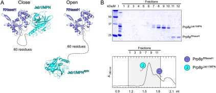
Figure 2.
Structure of Prp8pCTF* in isolation. (A) Two possible (close and open) configurations of Prp8pCTF*. (Blue) Prp8pRNaseH; (cyan) Prp8pJab1/MPN; (dashed line) flexible linker. Two crystallographically related Jab1/MPN domains are shown with respect to the same RNaseH domain. Other, primarily open, configurations in which the distances between the domains could be bridged by the linker can be found with other symmetry-equivalent Jab1/MPN domains. (B) Gel filtration analysis of Prp8pCTF* after treatment with trypsin. Details and labeling are as in Figure 1, C–I. After treatment with trypsin, the separated domains elute as isolated molecules, indicating that they do not stably interact in solution (cf. Fig. 1F).

