Figure 1.
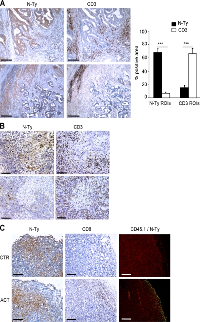
Chemical barriers inhibit T cell infiltration within the tumor. (A) Two representative serial sections of human colon carcinomas were immunostained (brown) for CD3 or nitrotyrosine, as indicated. Bars, 500 µm. Noncontiguous regions of interest (ROIs) with dimensions of 157 × 157 pixels were selected for either bright nitrotyrosine or CD3 staining and applied to serial sections. The percentages of immunoreactive areas for both nitrotyrosine and CD3 were obtained for each region of interest. ***, P < 0.001. Data are expressed as the means ± SE. (B) Two representative serial sections of human undifferentiated nasopharyngeal carcinomas were immunostained (brown) for CD3 or nitrotyrosine, as indicated. Bars, 200 µm. (C) CD45.1+ OT-I CTLs were adoptively transferred (ACT) to CD45.2+ mice bearing EG7-OVA tumors and, 4 d later, tumors were removed and stained to visualize nitrotyrosine-positive cells (N-Ty) and CTLs (either CD8+ or CD45.1+ cells). The first and second columns show the immunohistochemical detection of nitrotyrosine (N-Ty) and CD8+ T cells, respectively, whereas the third column shows immunofluorescence staining for CD45.1 (green) and N-Ty (red). Bars, 200 µm.

