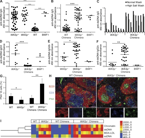
Figure 1.
WASp−/− B cells are sufficient for high-affinity, class-switched autoantibody production and spontaneous GC formation. IgG anti-dsDNA autoantibody ELISAs in 6.5–12-mo-old female WASp+/−, WASp−/−, and WT mice (A) and WT and WASp−/− chimeras at 16 wk after transplant (B). BWF1: lupus-prone positive control, NZB/NZW-F1, 8 mo old. Sera diluted 1:200; each dot represents an individual animal. (C) ELISAs with low versus high stringency washing conditions used to detect high-affinity IgG anti-dsDNA antibodies in WASp−/− and WASp−/− chimeric mice (12 wk old or 12 wk after transplant). Each pair of bars represents an individual animal. (D–F) ELISAs to detect IgG subclass-specific anti-dsDNA antibodies. (WASp−/− chimeras, n = 29; WASp−/− mice, n = 10; 4 mo after transplant or 4 mo old, respectively). (G) Percentage of splenic B cells staining positive for PNA by flow cytometry in 6–8-mo-old WT (n = 7) and WASp−/− (n = 7) mice, as well as WT (n = 5) and WASp−/− (n = 4) BM chimeras 6–8 mo after transplant. (H) Immunofluorescent staining of splenic sections from BM chimeras showing representative follicles using B220 (red) CD3 (blue) and PNA (green); 10× objective was used for image capture; bars, 100 µm. (I) Antigen microarray showing IgG reactivity of WT, WASp−/−, WT chimera, and WASp−/− chimera sera with ssDNA, dsDNA, chromatin, and MDA-LDL. Sera from WT and WASp−/− mice (1 yr) and chimeric mice (6 mo after transplant). Scale shows digital fluorescence intensity units; 600 represents threshold for reactivity as described in Materials and methods. These data are representative of 4 independent experiments with 20–30 mice per experiment. *, P < 0.05; **, P < 0.005; ***, P < 0.0005.

