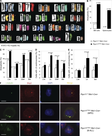
Figure 7.
Centrosome amplification and chromosomal instability in Ptpn11E76K/+ hematopoietic cells. (A) T-ALL tumor cells from pI-pC–treated Ptpn11E76K/+/Mx1-Cre+ mice were examined by spectral karyotype analyses. 12 metaphase spreads were examined for each sample. A representative result is shown. (B) BM cells and splenocytes freshly isolated from Ptpn11E76K/+/Mx1-Cre+ mice at the MPD stage and Ptpn11+/+/Mx1-Cre+ control mice (6–8 wk after pI-pC treatment) were assayed by karyotyping analyses. Cells (nonmegakaryocytes) with more or <40 chromosomes were counted as aneuploid cells. Results shown are a summary of the data obtained from five mice in each group. (C) LSK cells, LK cells, and GMPs were sorted from the BM of Ptpn11E76K/+/Mx1-Cre+ mice at the MPD stage and Ptpn11+/+/Mx1-Cre+ control mice. Purified cells, along with whole BM cells, were cultured in IMDM medium containing Tpo (20 ng/ml), FIt3 ligand (50 ng/ml), SCF (50 ng/ml), IL-3 (20 ng/ml), IL-6 (20 ng/ml), and 10% FBS (for LSK cells) or SCF (50 ng/ml), IL-3 (20 ng/ml), IL-6 (20 ng/ml), and 10% FBS (for other cell types) for 60 h (LSK and LK cells), 48 h (BM cells), and 16 h (GMPs) hours. Cells (nonmegakaryocytes) were then examined by karyotyping analyses, as described. (D) LSK cells, LK cells, and GMPs were sorted from the BM of Ptpn11E76K/+/Mx1-Cre+ mice at the MPD stage and Ptpn11+/+/Mx1-Cre+ control mice. Purified cells along with whole BM cells were immunostained with anti–γ-tubulin antibody. Centrosomes were then examined under a fluorescence microscope. For LSK cells, at least 100 cells were surveyed in each experiment. For the other cell types, at least 300 cells were examined in each experiment. Results shown are mean ± SEM of three independent experiments. (E) Purified LSK cells, LK cells, and unsorted BM cells were cultured in the medium (as described) for 48 h. Cells were then immunostained with anti–γ-tubulin antibody. Centrosomes were examined under a fluorescence microscope. Data shown are mean ± SEM of three independent experiments. (F) LK cells purified from Ptpn11E76K/+/Mx1-Cre+ mice at the MPD stage and Ptpn11+/+/Mx1-Cre+ control mice (top), and BM cells from Ptpn11E76K/+/Mx1-Cre+ mice with B-ALL (bottom) were immunostained with anti–γ-tubulin and Shp2 antibodies. Nuclei were counterstained with DAPI. The images were captured and analyzed using a laser-scanning confocal microscope. Bars, 5 µm.

