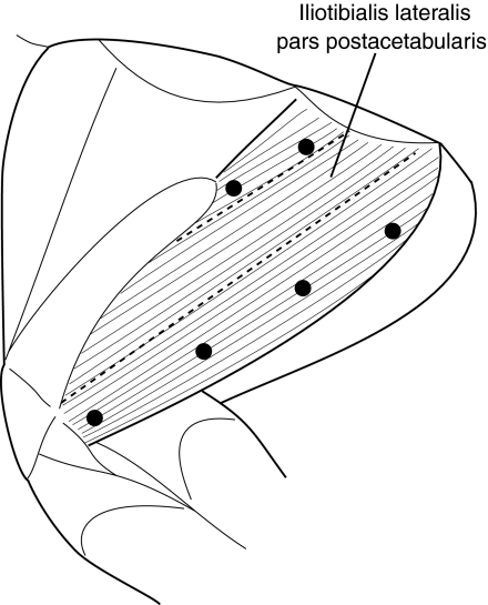
Fig. 1.
Position of the iliotibialis lateralis pars postacetabularis (ILPO) in the guinea fowl hindlimb. Black circles indicate the position of the sonomicrometry crystals. EMG electrodes were placed along the fascicles at the approximate mid-point of each pair of sonomicrometer transducers. Dashed lines indicate the divisions used to calculate the weight average strain and velocity. Figure drawn by Dr David Ellerby.

