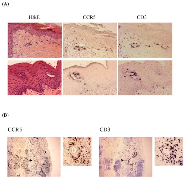
Figure 1. Infiltrating T cell lymphocytes in human aGVHD tissues are predominantly CCR5+.
(A) Skin and lip biopsies of GVHD patients. Three adjacent sections of skin (top panel) and lip (lower panel) biopsies of a representative GVHD patient were stained with H&E, CCR5 antibody or CD3 antibody. CCR5+ and CD3+ lymphocyte infiltrates were found in both the dermal and epidermal layers. (B) Lip biopsy of a representative GVHD patient with damaged salivary gland. Right and left panels are adjacent sections stained with CCR5 or CD3 antibodies. In each panel, the right side is an enlarged picture of the area with the arrow. CCR5+ and CD3+ lymphocyte infiltrates were found in the damaged area of salivary gland.

