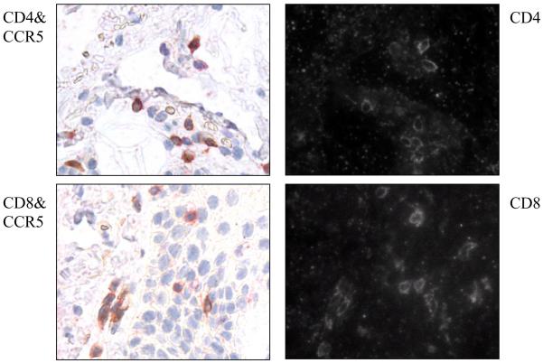
Figure 2. Infiltrating CCR5+ T cell lymphocytes in the skin of aGVHD are both CD4+ and CD8+ T cells.
The skin biopsies of a representative GVHD patient were double stained with CCR5 antibody and either CD4 antibody (top panel) or CD8 antibody (bottom panel). On the right side, CCR5+ cells are brown, and both CD4+ and CD8+ cells are Texas-Red. The same slides were photographed by light (right panel) and fluorescence microscopy (left panel) for Texas-Red.

