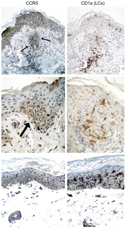
Figure 3. Strong CCR5 expression on T cells but not dendritic cells in aGVHD tissues.
Immunohistochemistry was performed on paraffin sections for CCR5 (left panel) and Langerhans DC marker CD1a (right panel). Top panel: skin biopsy with CCR5+ T cells in the superficial dermis (arrows) and epidermis in addition to CD1a+ Langerhans DCs at sites of lymphocytic infiltration. Middle panel: foci of epidermal damage with high-level CCR5 expression in T cells (arrow) but weak CCR5 staining in CD1a+ Langerhans DCs. Bottom panel: lip biopsy from a case with minimal epithelial damage and no detectable CCR5 expression in CD1a+ Langerhans DCs.

