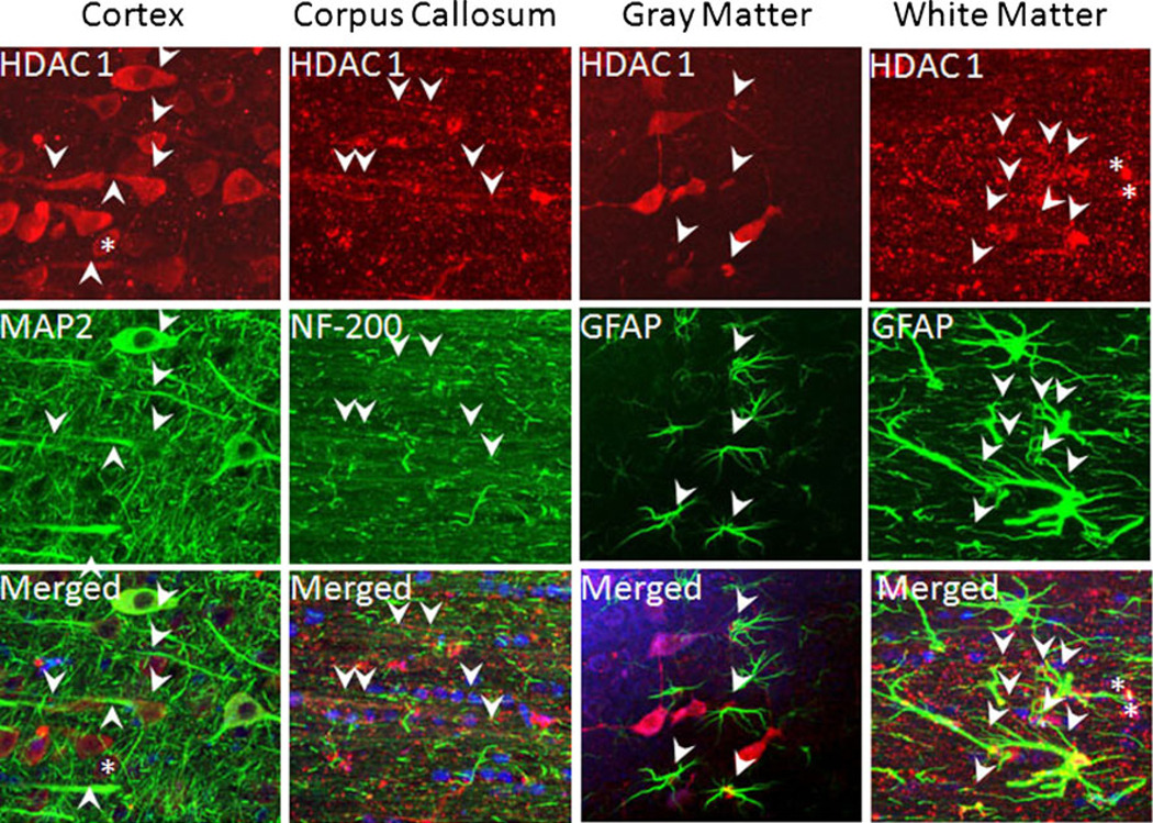
Fig. 2.
HDAC 1 expression in the cortex, corpus callosum, hippocampus, and subcortical white matter (SCWM). HDAC 1 displayed a clear cytoplasmic (arrows) and axonal (arrowheads) pattern in cortical neurons (cortex, upper panel) and co-localized with MAP2(+) neuronal cell bodies and axons (cortex, middle panel). Sytox(+) blue nuclei were visible in the merged images. Similarly, HDAC 1 (corpus callosum, upper panel) co-localized with NF-200(+) axons (arrowheads, left middle panel) in the corpus callosum. HDAC 1 was limited to the nuclei of GFAP(+) astrocytes in the hippocampus (gray matter, arrows). In SCWM, HDAC 1 labeling was detected in the nuclei (white matter, arrows) and to some extent in the proximal portions of the main processes and end-feet (arrowheads) of GFAP(+) astrocytes (green) outlining penetrating arterioles. Note the distinct co-localization of HDAC 1 and GFAP in the merged images, which confirmed that HDAC 1 exhibits a region (gray vs. white matter) specific labeling pattern in astrocytes

