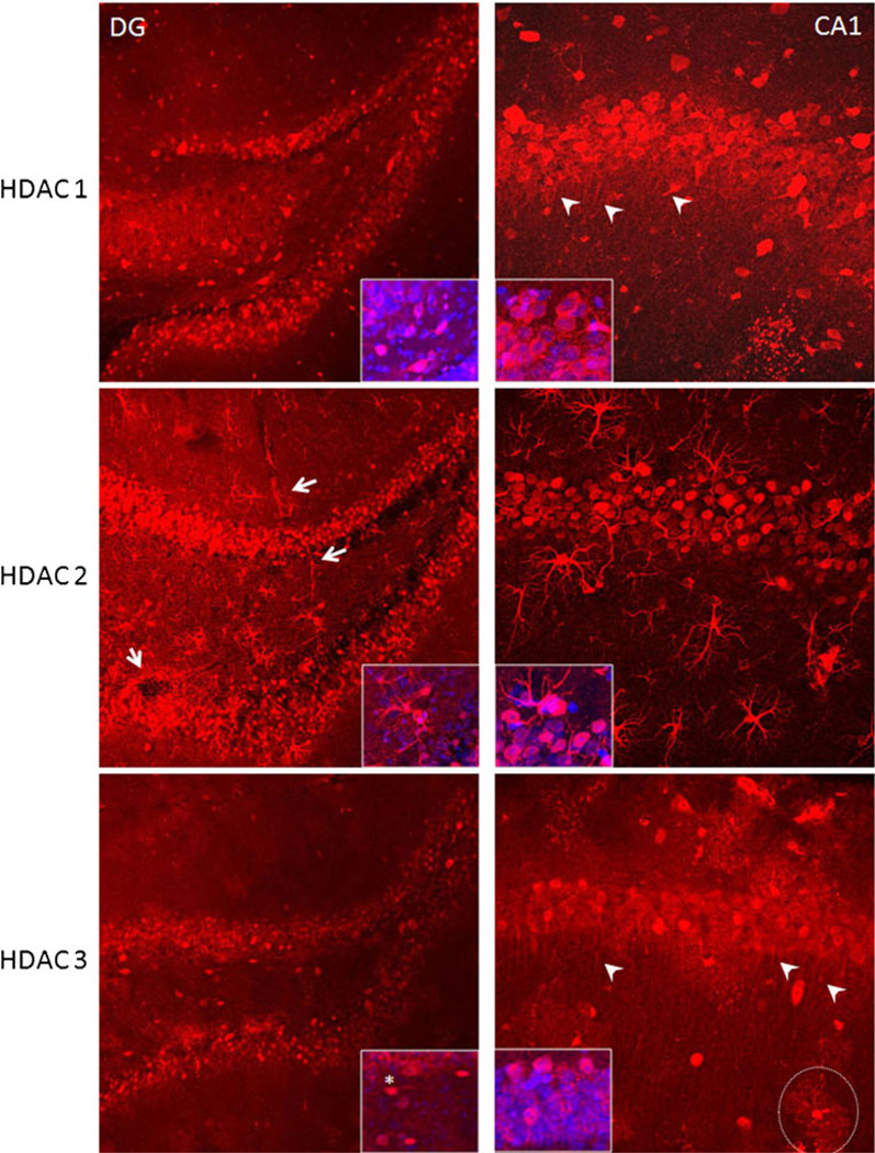
Fig. 5.
HDAC 1–3 expression in the dentate gyrus and the CA1 regions of hippocampus. HDAC 1 (upper panels) was compared in the dentate gyrus (DG), a site of neurogenesis, and the CA1 region which is known to undergo apoptosis even after very transient ischemic injury. HDAC 1 labeling was mainly expressed in the cytoplasm of DG granule cells and in CA1 pyramidal neurons. This was confirmed with blue Sytox(+) nuclei filling the center of neurons (see insets). Note that HDAC 1 labeled dendrites of CA1 pyramidal neurons (upper right panel, arrows), but not neuronal processes in DG. HDAC 2 (middle panels) was expressed in the nuclei of DG granule cells and in the cytoplasm of CA1 pyramidal cells (see insets for blue nuclei labeled with Sytox). In addition, astrocytes diffusely expressed HDAC 2 in their nuclei, cell bodies, processes, and end-feet outlining the vasculature (left middle panel, arrows). HDAC 3 was mainly expressed in the nuclei of DG granule cells and in the nuclei and cytoplasm of CA1 pyramidal cells. Note that some cells in between the upper and lower blades of the DG express HDAC 3 in their cytoplasm (insets). The punctate nature of HDAC 3 expression was more prominent in the CA1 region, especially around interneurons, and outlined their synaptic domains (dotted circle). HDAC 3 expression was also present in CA1 pyramidal cell dendrites (arrows)

