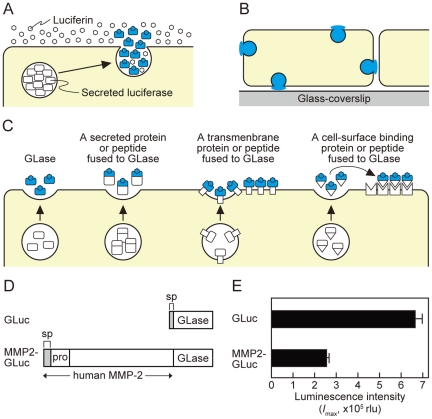
Figure 1. Bioluminescence imaging of GLase as a reporter protein to visualize proteins on the surface of mammalian cells.
(A) Schematic representation of imaging principles by GLase bioluminescence. (B) Detectable area of exocytosis in a single mammalian cell using GLase bioluminescence imaging method. (C) Schematic representation of the secretion of GLase and the fusion protein of GLase from mammalian cells and its binding on the cell surface. (D) Schematic representation of expression vectors for GLase and MMP2-GLase. GLuc (pcDNA3-GLuc); GLase with the signal peptide sequence, MMP2-GLuc (pcDNA3-hMMP2-GLuc); human MMP2 preproprotein fused to GLase. (E) GLase activity in the conditioned medium of HeLa cells. HeLa cells were transfected with pcDNA3-GLuc or pcDNA3-hMMP2-GLuc and cultured for 24 hr followed by incubation with HBSS for 60 min at 37°C. The luminescence activity in the conditioned medium was determined using a luminometer.

