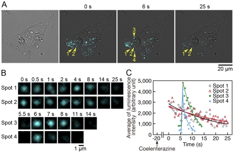
Figure 5. Identification of MMP2-GLase being secreted and bound on the cell surface of a HeLa cell using bioluminescence imaging.
Bioluminescence imaging of HeLa cells transiently expressing MMP2-GLase. Objective lens; 40×. Luminescence signals recording was started after 20 s from the addition of HBSS buffer containing coelenterazine with an exposure time of 500 ms for 75 s (Video image is in Video S4). (A) The bright-field image (the left panel), and luminescence images acquire at 0, 6, and 25 s. Two continuous luminescence spots (spot 1 and 2) are indicated by yellow arrowheads labeled with 1 and 2, respectively, and two transient diffusive luminescence spots (spot 3 and 4) are indicated by yellow arrowheads labeled with 3 and 4, respectively. (B) Magnified luminescence images of spot 1–4 indicated by arrowheads labeled with 1–4 in (A). (C) Time-dependent changes of the average luminescence intensity of spot 1–4 indicated by arrowheads labeled with 1–4 in (A).

