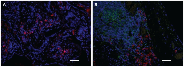
Figure 3. FLC-positive cells, B cells, and plasma cells in the lungs of HP and IPF subjects.
Representative pictures are shown of numerous FLC-positive cells (both kappa and lambda FLC are stained red) (A), and B cells (green) and plasma cells (red) (B) which are found in HP and IPF lung tissue. Scale bar: 50 µm.

