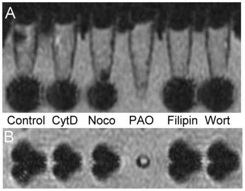
Figure 4. MRI of macrophages treated with different endocytotic inhibitors prior to SPIO particle uptake.
(A) Coronal view and (B) axial view under T2 weighted scanning protocol. Cells were centrifuged to the bottom of the test tube and imaged as dark signals. The coronal view and axial view of these cells are consistent. There was a drop in signal intensity in the cells treated with Ferucarbotran and inhibitors, including cytochalasin D (CytD), nocodazole (Noco), filipin, and wortmannin (Wort). In contrast, the signal intensity of the cells treated with phenylarsine oxide (PAO) revealed no loss in signal intensity.

