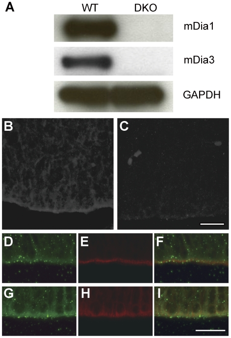
Figure 1. Expression of mDia1 and mDia3 in embryonic mouse forebrain.
(A) Western blotting for protein expression of mDia1 and mDia3 in the mouse forebrain at embryonic day 16 (E16). The specificity of mDia1 and mDia3 signals was confirmed using lysates from mDia-DKO mice. GAPDH was used as an internal control. (B, C) Immunofluorescent staining for mDia3 in coronal brain sections from wild-type (B) and mDia3null (C) embryos at E16. Note that mDia3 signals were enriched at the apical surface of the cerebral cortex in wild-type mice. (D–F) Immunofluorescent staining for mDia3 (green, D) and phalloidin staining (red, E) in coronal sections of the lateral ventrical wall from wild-type mice at E13. A merged image is shown in F. mDia3 accumulated at the apical surface and colocalized with filamentous actin. (G–I) Double immunofluorescent staining for mDia3 (green, G) and β-catenin (red, I) in coronal sections of the cerebral cortex from wild-type mice at E13. A merged image is shown in I. mDia3 also colocalized with β-catenin at the apical surface. (B, C) Scale bar, 20 µm. (D–I) Scale bar, 20 µm.

