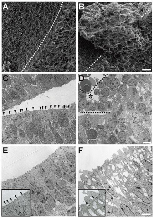
Figure 4. Widespread disruption of neuroepithelium architecture in mDia-DKO mice.
(A, B) Scanning electron micrograph of the surface of the lateral ventricle wall from wild-type (A) and mDia-DKO (B) mice at E16. Note that neuroepithelial cells protruded into the lateral ventricle in mDia-DKO mice. Dotted line indicates the cut edge of the ventricle wall. (C–F) Transmission electron micrograph of neuroepithelial cells lining the ventricle wall from wild-type (C, D) and mDia-DKO (E, F) mice at E16. An asterisk indicates a region of periventricular dysplastic mass marked by dotted lines. Arrowheads indicate apical adherens junction. Insets in E and F show higher magnification of the apical surface of neuroepithelium. (A, B) Scale bar, 10 µm. (C, D) Scale bar, 5 µm. (E, F) Scale bar, 5 µm.

