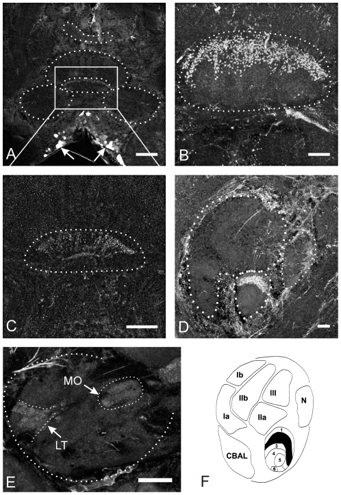
Figure 5. Nitric oxide-stimulated cGMP immunoreactivity in the central complex.
A: Frontal section, through the central complex and surrounding midbrain structures. Within the central complex cGMP-ir is restricted to a particular layer of the CBL. Labeled cell bodies are located in the inferior median protocerebrum (indicated by arrows). B and C: Frontal sections through the CBL. Strong cGMP-ir is associated with fibers of tangential neurons projecting close to the anterior border of the CBL. The neurites enter the CBL from posterior direction and innervate the CBL in a fan-shaped fashion (best seen in C). Immunostaining in the CBL appears to be beaded (best seen in B and D) suggesting that labeled neurites represent presynaptic structures. D: Sagittal section through the central body: Staining in the CBL is restricted to layer II, while the other layers of the CBL and the entire CBU are completely devoid of staining (compare with F). E: Frontal section through one lateral accessory lobe (LAL) containing faint labeling in the lateral triangle (LT) and the median olive (MO). F: Schematic drawing of a sagittal section through the CB (modified from [18], [21]. Regions labeled black contain cGMP immunopositive neurites. CBAL = anterior lip of the central body, N = noduli. Scale bars = 100 µm in A; 20 µm in B, D and E; 10 µm in C.

