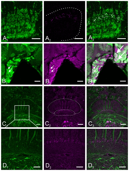
Figure 6. GABA, mAChR and NO-stimulated cGMP immunoreactivity in the central complex.
A1–B3 Double labeling of GABA (green) and cGMP (magenta) in frontal sections through the midbrain. Colocalization (highlighted in white) was detected in tangential neurons of the central body lower division. All cGMP-immunoreactive neurites also contain GABA immunoreactivity, whereas only a subset of GABA-immunoreactive neurites accumulated detectable amounts of cGMP upon nitric oxide stimulation. Colocalisation of GABA and cGMP immunoreactivity was detected in five cell bodies per hemisphere in the infero-median protocerebrum (two of them are marked by arrows in B1–B3). C1–D3 Double labeling of mAChR (green) and cGMP (magenta) on frontal sections through the central complex. No colocalisation was detected, indicating that NO has no direct effect on mAChR-expressing columnar neurons of the central complex. Scale bars = 50 µm in B1–C3; 20 µm D1–D3; 10 µm in A1–A3.

