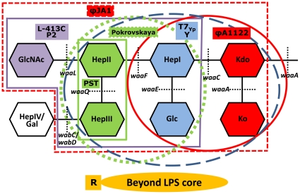
Figure 2. Identification of Y. pestis cell surface receptors for different bacteriophages.
The structure of LPS core is presented according to [51]. Kdo, 2-keto-3-deoxy-octulosonic acid; Ko, d-glycero-d-talo-oct-2-ulosonic acid; Hep, heptulose (ketoheptose); Glc, glucose; Gal, galactose; GlcNAc, N-acetylglucosamine. Black dashed lines designate the sites where the gene product, a corresponding glycosyltransferase, forms a glycosidic bond. The yrbH mutations affecting Kdo synthesis have the same effect on the LPS structure as waaA mutations, and defect in the hldE gene involved in ADP-l,d-heptose synthesis causes the same phenotype as the waaC mutation [51]. The critical sugar residues for certain phage receptors have solid color fill matching with that of phage designations and with the color of lines outlining the receptors. The phage receptors are outlined with bold purple (for the L-413C and P2 phages), dashed red (for ϕJA1), bold green (for PST), dashed green (for Pokrovskaya), dashed blue (for T7Yp and Y), and bold red (for ϕA1122) lines.

