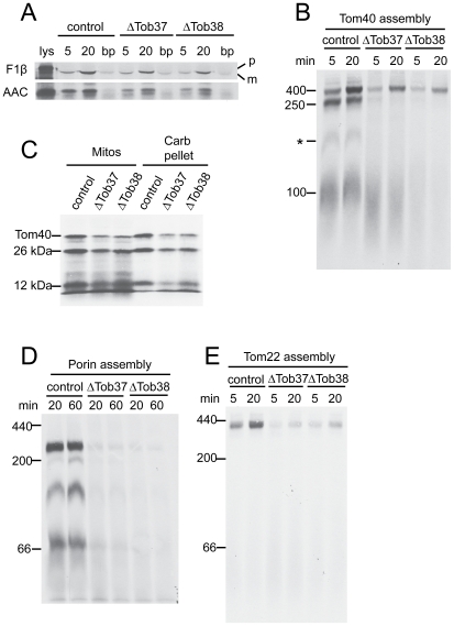
Figure 2. Import/assembly of mitochondrial precursor proteins into mitochondria deficient in Tob37 or Tob38.
(a) Radiolabeled matrix precursor F1β and inner membrane precursor AAC were incubated (for 5 min or 20 min, as indicated) with mitochondria isolated from heterokaryotic strains (indicated at the top of the panel) grown in the presence of histidine and fpa to reduce levels of Tob37 or Tob38 in the respective mutants. Following import, mitochondria were subjected to SDS-PAGE. Proteins were transferred to nitrocellulose membrane, and import was analyzed by autoradiography. (control, strain HP1; lys, 33% of the radiolabeled lysate added to each import reaction; bp, “bypass import” in mitochondria treated with trypsin to remove surface receptors prior to the import reaction; p, precursor protein; m, mature protein.) (b) Radiolabelled Tom40 precursor was incubated for 5 min and 20 min with mitochondria isolated from the strains indicated (top of panel) grown in the presence of histidine and fpa. Mitochondria were dissolved in 1% digitonin and subjected to BNGE. The proteins were transferred to PVDF membrane and analyzed by autoradiography. The size of the mature TOM complex (400 kDa), and assembly intermediates I (250 kDa) and II (100 kDa) are indicated on the left. * indicates an undefined band. (c) Tom40 was imported into mitochondria isolated from the strains indicated for 20 min. Following import, proteinase K was added to each import reaction for 15 min on ice. PMSF was added to inactivate the proteinase, each reaction was divided into equal halves, and mitochondria were pelleted. One half was suspended in SDS-PAGE cracking buffer (Mitos). The other half was suspended in sodium carbonate (pH 11.5) and incubated on ice for 30 min. The membrane sheets were pelleted and suspended in cracking buffer (Carb pellet). Both sets of reactions were subjected to SDS-PAGE and the proteins were transferred to nitrocellulose membrane and examined by autoradiography. The positions of Tom40 and the 26 kDa and 12 kDa fragments generated by proteinase K digestion are indicated. (d) As in panel B except that mitochondria were incubated with the radiolabeled precursor of porin. The numbers on the left indicate the position of molecular weight markers. (e) Assembly of Tom22. As in panel D, except mitochondria were incubated with radiolabeled Tom22.

